
Fig. 4.
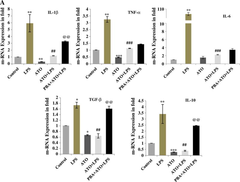
Diminished cytokine production in ATO-treated Raw 264.7 cells was dependent on UPR signaling: Cytokine production was quantified by real time quantitative PCR (qRT-PCR) using SYBR Green methodology. Transcription levels of IL-1β, TNF-α, IL-6, TGF-β, and IL-10 were determined and presented in terms of fold change. (A) Cells received five different treatments. (1) Control-treated with normal saline, (2) LPS-treated with 250 ng/ml for 3 h, (3) ATO-treated with 2 μM for 3 h, (4) pre-treated with ATO followed by LPS, and (5) pretreated with PBA (1 mM for 24 h) followed by ATO and LPS. ATO-treatment to murine macrophage reduced the cytokine production alone as well as in combination of LPS stimulation. PBA pretreatment to the macrophage restored the cytokine production of ATO and LPS-induced cytokine production. Data are expressed as Mean ± SE of at least three independent samples. Statistical significance was determined using Student’s t test. #,*P > 0.05, ##,**P > 0.01 and ###,***P > 0.001 show significance levels. * indicates significant level when compared to saline-treated control cells. # indicates significant level when compared to LPS-treated cells. @ indicates significant level when compared to ATO + LPS-treated cells.