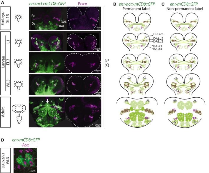
Left: Schematics of CNSs of various stages with dotted outlines indicating central brain regions indicated in (B). Right: Time course pictures of brains containing permanently labeled cells in the En expression domain co‐stained for Poxn (maximum intensity projections; split magenta channel). At embryonic stage 15, clusters of en cells in the protocerebrum (Pc), deutocerebrum (Dc), and tritocerebrum (Tc) can be detected, which have been named in antero‐posterior order: (i) P/PC/b1/DALv (for dorso‐antero‐lateral)/MC (for medial cluster—because of later emergence of a cluster anterior to this one—see below); (ii) D/DC/b2/BAla (for baso‐antero‐lateral)/PC (for posterior cluster, a nomenclature which could be confused with that for the protocerebral cluster); and (iii) T/TC/b. In first‐instar larvae (L1), an additional protocerebral cell cluster is visible antero‐dorsal to the DALv (arrowhead), which starts expressing en after embryonic stage 15, and that has been named DPLam (for dorso‐postero‐lateral)/AC (for anterior cluster). Asterisks, neuropil structures; arrow, ellipsoid body of the central complex (adult structure).

