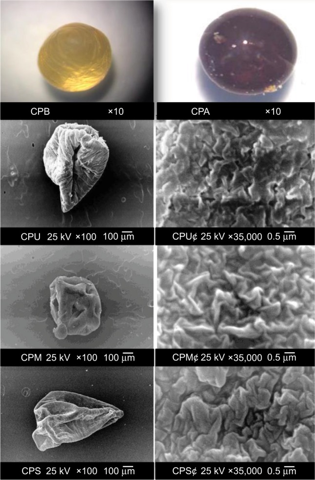
Figure 2.

SEM images of loaded and unloaded capsules.
Notes: Stereo optical microscope morphology of whole shape (magnification, ×10) of the capsules prepared from chitosan (1%), alginate (1%), glutaraldehyde (2%), and gelatin (2.5%), before (CPB) and after (CPA) drying. SEM photograph of unloaded capsules (CPU) and their surface morphology (CPU¢), capsules loaded with malathion (CPM) and their surface morphology (CPM¢), and capsules loaded with spinosad (CPS) and their surface morphology (CPS¢). Scale bar is 100 μm and magnification is ×100 for whole capsules, and scale bar is 0.5 μm and magnification is ×35,000 for their surface morphologies.
Abbreviation: SEM, scanning electron microscopy.
