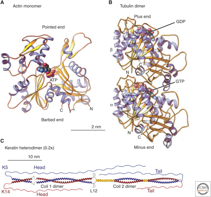
Figure 2.
Structures of (A) an actin monomer, (B) a tubulin dimer, and (C) a keratin heterodimer. Ribbon diagrams compare the building blocks of the three cytoskeletal polymers. The scale of the keratin is 20% that of actin and tubulin to fit this long molecule into the figure. (A,B, Reprinted from Pollard and Earnshaw 2008; C, reprinted from Herrmann and Aebi 2016.)

