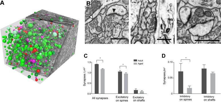
Fig 2. Synapse density decreases per unit volume in adult and aged mouse layer 1 neuropil.
A, To count synapses in stacks of serial FIBSEM images (3 adult, and 3 aged), circles were placed at the center of each synapse using the TrakEM2 software in FIJI (www.fiji.sc); with their diameter equal to the maximum diameter of each contact. B, micrographs showing: left, an asymmetric synapse on a spine (presumed glutamatergic, black arrowhead) and a symmetric synapse on a shaft (presumed inhibitory, white arrowhead); center, an asymmetric synapse on a shaft; right, a multi-synaptic bouton (MSB). C, The density of all synapses was significantly lower in aged mice (adult, 1.40 ± 0.006 per μm3; aged, 1.17 ± 0.013 per μm3; t-test, *, p < 0.001) with a significant drop in asymmetric synapses on dendritic spines (adult, 1.05 ± 0.03 per μm3; aged, 0.96 ± 0.02 per μm3; t-test, *, p < 0.001). On dendritic shafts, there were less asymmetric synapses, but the drop was not significant (adult, 0.17 ± 0.03 per μm3; aged, 0.11 ± 0.02 perμm3; t-test, p = 0.09). D, Symmetric (presumed inhibitory) synapses were also significantly reduced, but only on dendritic spines (adult, 0.07 ± 0.01 per μm3; aged, 0.02 ± 0.006 per μm3; t-test, *, p = 0.0028) and not on shafts (adult, 0.08 ± 0.07 per μm3; aged, 0.065 ± 0.003 per μm3; t-test, p = 0.27). Scale bar in B is 500 nanometers.

