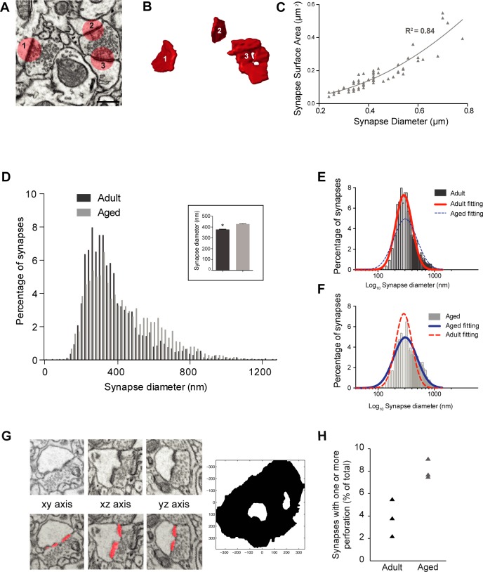
Fig 3. Synapses are larger in aged layer 1 neuropil.
A, FIBSEM electron micrograph shows two boutons making asymmetric synapses; synapse 1 is made with a single synaptic bouton, 2 and 3 with a multi-synaptic bouton. B, the same synapses were segmented in the TrakEM2 software in FIJI (shown in red: www.fiji.sc) and reconstructed in 3D. C, There is a strong correlation between the diameter of circles used to annotate 57 synapses and their surface areas measured from their 3D reconstruction (slope of second polynomial regression, y = 0.032–0.09x + 0.95x2; R2 = 0.84). D, Frequency distribution of asymmetric synapse sizes, estimated from the maximum diameter measurements shown in Fig 2 (Inset shows average values of all measurements, N = 2800 adult synapses, 377.8 ± 3.2 nm; N = 2423 aged synapses, 427.7 ± 3.9 nm; p < 0.0001, Kolmogorov-Smirnov test), showed that synapses in layer 1 of aged mice are larger. E, The same data plotted on a logarithmic scale for adult, and F, aged mice. Both show a log-normal distribution (adult, red fitting; center = 287.5, amplitude = 7.27, width = 0.33, R2 = 0.96; aged, blue fitting; center = 305.1, amplitude = 4.9, width = 0.47, R2 = 0.94). G, Examples of a series of micrographs containing a perforated synaptic density (top) and its segmentation (bottom) by means of the automated detection method, and its 3D rendering on the right. H, Graph showing the percentage of asymmetric synapses that displayed one or more perforation.

