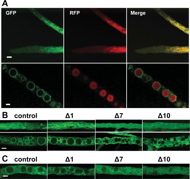
Fig 7. Subcellular localization of NCU05950 protein.
(A) A heterokaryon expressing both a GFP fusion protein of NCU05950 and an RFP fusion protein of the vacuolar protein VAM-3 was observed in living hyphae by confocal microscopy. Top row: tips of growing hyphae. Bottom row: older hyphae containing large round vacuoles. Left column: green channel, GFP. Middle column: red channel, RFP. Right column: merged channel, red + green. Scale bars = 5 μm. (B) Subcellular localization of NCU05950 N-terminal deletion mutants. The GFP fusion protein of NCU05950 expressed from the ccg-1 promoter was modified by deleting either 1, 7 or 10 amino acids after the initial methionine at the N-terminus (indicated as Δ1, Δ7 and Δ10). GFP localization was observed in living hyphae by confocal microscopy. Upper panels show young hyphae. Lower panels show older hyphae with mature round vacuoles. Control = full-length NCU05950. Scale bar = 5 μm. (C) Subcellular localization of GFP fusion proteins driven by the native promoter. The GFP fusion protein of NCU05950 was expressed from the NCU05950 promoter. N-terminal deletions and microscopy as for (B). Older hyphae are shown. Control = full-length NCU05950. Scale bar = 5 μm.

