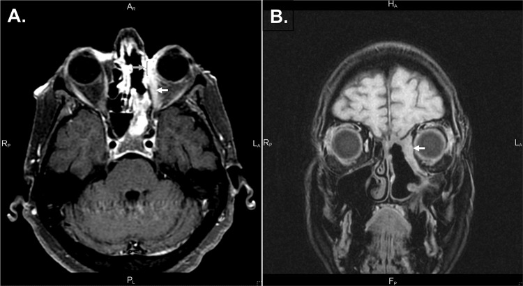
Figure 2.
T1-weighted magnetic resonance imaging with contrast, fat saturation sequence. (A) T1 fat saturation with contrast; dehiscence of the left lamina papyracea (gray arrow and bracket); enlargement of medial left medial rectus along the entire length of muscle (white arrow). (B) Thickening of the left medial orbital wall, obscuring the medial rectus.

