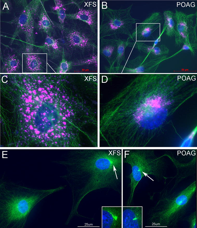
Figure 3. Salient phenotypic differences between TF derived from XFG and POAG donors.

XFG and POAG TF were seeded under serum-free (starvation) conditions. A,B. Distribution of microtubules (β-tubulin, green) and lysosomes (LAMP1, pink), Bar=50um. C,D Magnified images. In XFG cells, with the exception of rare cells, the β-tubulin stain remains outside the nucleus and the lysosomes are minimally condensed. In the POAG cells lysosomes and β-tubulin staining is highly concentrated at a typical MTOC structure. E,F. In XFG cells the concentration of γ-tubulin remains outside the nucleus and is either small of amorphous γ-tubulin staining (arrow). In POAG cells, a large fraction of the stain is localized in a tubular or conical small structure consistent with the γ-TurC morphology that clearly penetrates deep into the nucleus (arrow). Bar = 25um. N=3.
