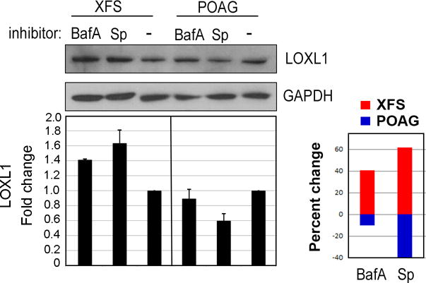
Figure 4. Effect of Bafilomycin-A1 and Spautin-1 on LOXL1 protein.

XFS and POAG TF were seeded in serum-free (starvation) conditions for 18 hours with 0.5uM Bafilomycin-A1 (BafA), 10uM Spautin-1 (Sp) or in control medium, without inhibitors. Western blot for LOXL1 shows that in XFG cells the amount of LOXL1 increases after treatment with autophagy inhibitors, whereas in POAG cells these inhibitors reduce LOXL1 amounts. Loading control, GAPDH. Statistical results for N = 4 are shown as the percent change (up or down) for XFG and POAG cells with each inhibitor.
