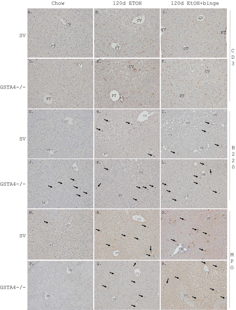
Figure 1. Increased inflammation following 116d chronic EtOH consumption +/− Binge.

Formalin fixed tissue sections were analyzed immunohistochemically using polyclonal or monoclonal antibodies directed against Panels A-C SV CD3 chow, 116d EtOH, 116d EtOH +Binge, Panels D-F GSTA4−/− CD3 chow, 116d EtOH, 116d EtOH +Binge), Panels G-I SV B220 chow, 116d EtOH, 116d EtOH +Binge, Panels J-L GSTA4−/− B220 chow, 116d EtOH, 116d EtOH +Binge, Panels M-O SV MPO chow, 116d EtOH, 116d EtOH +Binge, P-R GSTA4−/− MPO chow, 116d EtOH, 116d EtOH +Binge. Arrows indicate B220 and MPO positive cells. Representative images are shown; n = at least 3 mice per genotype (CV, central vein, PT, portal triad).
