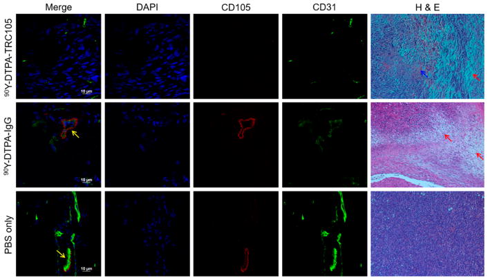
Figure 4.
Immunofluorescent and H&E staining of tumor tissues. Colocalization of CD31 and CD105, indicated by yellow arrows, shows angiogenic vessels. H&E staining visualized notable damage to 90Y-treated tumors, with red arrows pointing out representative damaged areas, and blue arrows indicating hemorrhaging.

