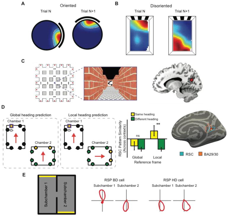
Figure 3. Anchoring the cognitive map to the world.
A) In oriented rats, from trial-to-trial, the orientation of the hippocampal map is set by featural cues on the walls of the chamber, rotating in concert with rotation of those cues. B) Following disorientation, the hippocampal map is anchored primarily by the geometric shape of the chamber rather than featural cues. For this example place cell, from trial-to-trial, two place fields were observed relative to chamber geometry, one being 180° rotation of the other, mirroring the chamber’s geometric symmetry (adapted from ref. 64). C) fMRI evidence that human retrosplenial/medial parietal region represents heading direction (adapted from ref. 87). During scanning, participants were shown pictures associated with different facing directions learned in a virtual-reality arena (left). fMRI adaptation was found in medial parietal cortex (BA 31) when the same facing direction was elicited on successive trials (right). D) fMRI evidence that the retrosplenial complex (RSC) represents heading in a local reference frame (adapted from ref. 85). During training before scanning, participants learned the locations of objects (denoted by circles) inside virtual reality museums. During scanning, participants performed a task that required them to imagine facing each object encountered during training. Multivoxel activity patterns in RSC were similar for facing directions across the two museums defined in a local, but not global, reference frame. E) In rodents, retrosplenial cortex (RSP) contains both “bidirectional” (BD) cells that represent heading in a local reference frame and head direction (HD) cells that represent heading in a global reference frame (adapted from ref. 95).

