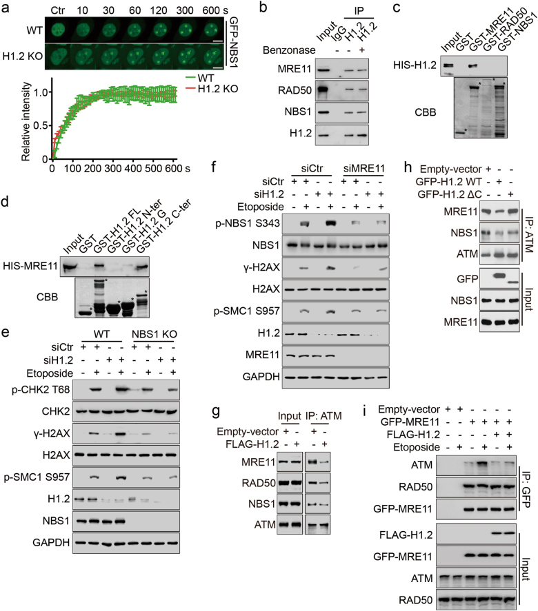
Fig. 3.
Linker histone H1.2 inhibits ATM recruitment and activation by interacting with MRN. a Wild type and H1.2 KO (1#) HeLa cells were transfected with GFP-NBS1 and subjected to laser micro-irradiation-coupled live-cell imaging. Images were taken every 10 s for 10 min and the relative intensity of the irradiation path signal was shown. The data represent the mean ± SD. Scale bars, 10 μm. b HeLa cells extracts were analyzed by Co-IP assay with or without benzonase treatment with the indicated antibodies. c GST alone or GST-MRE11, RAD50 and NBS1 were incubated with HIS-H1.2 for GST pull-down assay. * indicates specific protein bands. d GST alone or GST-H1.2 fragments were incubated with HIS-MRE11 for GST pull-down assay. * indicates specific protein bands. e Wild type or NBS1 KO HeLa cells were transfected with the indicated siRNAs and treated with 40 μM etoposide for 2 h and analyzed by immunoblotting. f HeLa cells were transfected with the indicated siRNAs and treated with 40 μM etoposide for 2 h and analyzed by immunoblotting. g, h HeLa cells were transfected with the indicated plasmids, and the whole cell lysates were immunoprecipitated with ATM antibody and analyzed by immunoblotting. i HeLa cells were transfected with the indicated plasmids and treated with 40 μM etoposide for 2 h. Whole cell extracts were prepared and analyzed by Co-IP assay and immunoblotting with the indicated antibodies

