Fig. 4.
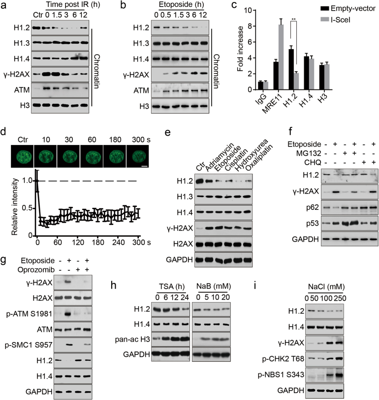
Linker histone H1.2 is rapidly displaced from chromatin and degraded upon DNA damage. a, b HeLa cells were exposed to 10 Gy irradiation (IR) and released at the indicated time or treated with 20 μM etoposide for the indicated time. Chromatin was fractionated and subjected to immunoblotting. c DR-GFP U2OS cells were transfected with I-SceI endonuclease or an empty vector and subjected to chromatin immunoprecipitation with the indicated antibodies followed by real-time PCR 48 h post transfection. All data represent the mean ± SD. d HeLa cells were transfected with GFP-H1.2 and subjected to laser micro-irradiation-coupled live-cell imaging. The initial signal intensity of GFP-H1.2 was normalized to 1 and ~15 IR paths from 10 separate cells were calculated. All data represent the mean ± SD. Scale bars, 10 μm. e HeLa cells were treated with either 1 μM adriamycin at 1 μM, 20 μM etoposide, 10 μM cisplatin, 2 mM hydroxyurea or 10 μM oxaliplatin for 12 h and analyzed by immunoblotting. f HeLa cells were treated with 1 μM MG132 or 50 μM CHQ for 12 h with or without 20 μM etoposide and analyzed by immunoblotting. g HeLa cells were treated with etoposide at 20 μM for 12 h with or without Oprozomib at 100 nM and analyzed by immunoblotting. h HeLa cells were treated with 1 μM TSA for the indicated time or to increasing concentrations of sodium butyrate (NaB) for 12 h and analyzed by immunoblotting. A pan-ac H3 antibodies was used as a positive control. i HeLa cells were exposed to increasing concentrations of sodium chloride (NaCl) for 2 h and analyzed by immunoblotting

