Figure 1.

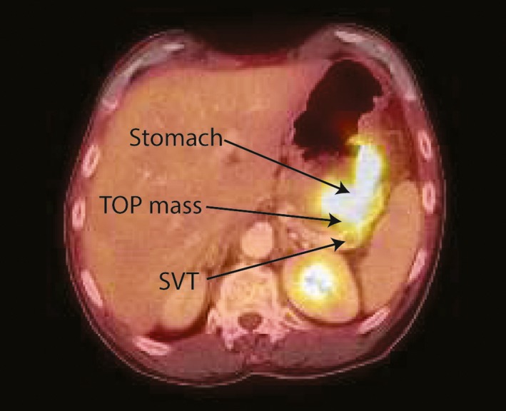
PET‐CT image showing thickening of the wall of the gastric fundus, a mass in the tail of the pancreas (both with increased radiotracer uptake), and a 1.1‐cm filling defect within the splenic vein

PET‐CT image showing thickening of the wall of the gastric fundus, a mass in the tail of the pancreas (both with increased radiotracer uptake), and a 1.1‐cm filling defect within the splenic vein