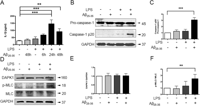
Figure 1.
Aβ25–35 induced caspase-1 and DAPK1 activation in LPS-primed Bv2 cells. (A) Cells were primed with LPS (100 ng/ml) for 6 h, and treated with Aβ25–35 (25 μM) for varying times (6 h, 24 h, 48 h); the amount of IL-1β in the culture supernatant was assessed by ELISA. (B–F) Cells were primed with LPS and stimulated with Aβ25–35 for 24 h. Protein levels of caspase-1 (B–C), DAPK1 and p-MLC (D–F), see original blots in Supplementary Fig. S2) were assessed by western blotting analysis. Data are shown as mean ± SEM for three independent experiments. **P < 0.01, ***P < 0.001. Aβ: β-amyloid; DAPK1: death-associated protein kinase 1; LPS: lipopolysaccharide; MLC: myosin II regulatory light chain.

