Fig. 1.
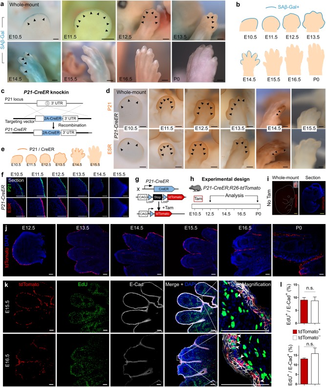
Embryonic senescent cells re-enter cell cycle and contribute to tissues after birth. a Whole-mount SAβ-Gal staining on forelimbs of E10.5–P0 mice. Arrowheads indicate SAβ-Gal+ cells. b Cartoon image showing SAβ-Gal activity pattern. No SAβ-Gal+ cell is detected after E15.5. c Generation of P21-CreER knock-in allele. d Whole-mount immunostaining for P21 or ESR on P21-CreER embryos. e Cartoon image showing expression pattern of P21 and CreER on P21-CreER mouse limbs. f Immunostaining for P21 and ESR on P21-CreER limb sections. g Strategy for genetic lineage tracing by tamoxifen (Tam)-mediated Cre-loxP recombination. h Schematic figure showing experimental strategy. Tam tamoxifen. i Whole-mount and sectional view of tdTomato expression in P21-CreER;R26-tdTomato embryo without tamoxifen (No Tam) treatment. j Immunostaining for tdTomato on E12.5–P0 mouse limb sections. tdTomato+ cells persist after birth. k Immunostaining for tdTomato, EdU, and E-cadherin (E-Cad) on E15.5 and E16.5 limb sections. Arrowheads indicate proliferating tdTomato+ cells. l Quantification of the percentage of proliferating tdTomato+ epithelial cells. n = 5; n.s., nonsignificant. Scale bars: yellow, 1 mm; black, 200 µm; white, 100 µm. Each figure is representative of five individual samples

