Figure 2.
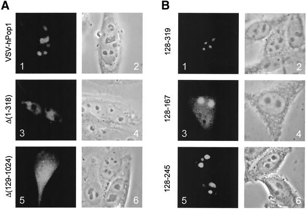
Subcellular localization of deletion mutants of hPop1. (A) VSV-hPop1 constructs were transiently transfected into HEp-2 cells. Cells were fixed with paraformaldehyde, and the expressed proteins were visualized with the use of anti-VSV antibodies (panels 1, 3, and 5). A phase-contrast image of the same cells is shown in panels 2, 4, and 6. Panels 1 and 2: VSV-hPop1 construct; panels 3 and 4: VSV-hPop1 Δ(1–318); panels 5 and 6: VSV-hPop1 Δ(129–1024). (B) EGFP-hPop1 constructs were transiently transfected into HEp-2 cells. Cells were fixed with methanol/acetone, and the fluorescent proteins were visualized by direct fluorescence microscopy (panels 1, 3, and 5). Phase-contrast images of the same cells are shown in panels 2, 4, and 6. Panels 1 and 2: EGFP-hPop1 128–319; panels 3 and 4: EGFP-hPop1 128–167; panels 5 and 6: EGFP-hPop1 168–245.

