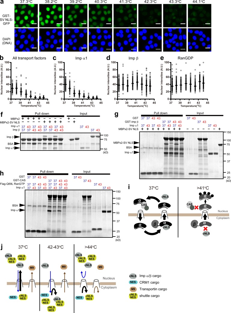
Figure 5.
Thermosensitivity of Imp α1. (a and b) Using Imp α1, Imp β, and RanGDP preincubated at indicated temperatures, in vitro transport assays of GST-SV NLS–GFP were performed. After the reactions, the cells were fixed and stained with DAPI (a). Then, nuclear intensities of GST-SV NLS–GFP were plotted (b). Bars, 20 µm. (c–e) Imp α1 (c), Imp β (d), or RanGDP (e) was preincubated for 60 min at indicated temperatures, and the remaining two transport factors were preincubated at 37.3°C. Then, the import assays of GST-SV NLS–GFP were performed. The nuclear intensities were plotted (see also Fig. S3, a–c). Black bars show the median of >100 cells at eight temperature conditions (total n = 1,521 [b], 1,255 [c], 1,598 [d], and 1,494 [e]). All plots in each graph were calculated from one simultaneous experiment. (f) Imp α1 and Imp β were independently preincubated at either 37.3 or 43.3°C for 60 min. Then, these proteins and either MBPx2 or MBPx2-SV NLS were incubated with amylose beads. (g) GST, GST–Imp β, and Imp α1 were independently preincubated at either 37.3 or 43.3°C. Then, either GST or GST–Imp β was incubated with Imp α1, MBPx2-SV NLS, and glutathione beads. (h) GST, GST-CAS, Flag-RanGTP (Q69L), and Imp α1 were independently preincubated at either 37.3 or 43.3°C. Then, either GST or GST-CAS was incubated with Imp α1, Flag-RanGTP (Q69L), and glutathione beads. Proteins bound to beads were analyzed by Coomassie brilliant blue staining. (i) Model for the inhibition of Imp α/β–dependent import and the nuclear translocation of Imp α. (j) Model for temperature-dependent transport modulations. cNLS indicates classical NLS, which is recognized by Imp α1.

