Figure 1.
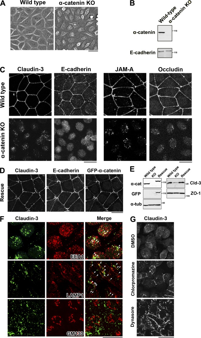
α-Catenin–KO cells internalize claudins. (A) Phase-contrast images of WT and α-catenin–KO EpH4 cells. (B) Immunoblotting of whole-cell lysates of WT and α-catenin–KO EpH4 cells with the indicated antibodies. (C) WT and α-catenin–KO EpH4 cells were fixed and costained with an anti–claudin-3 pAb and an anti–E-cadherin mAb (left) or with an anti–JAM-A pAb and an antioccludin mAb (right). (D) α-Catenin–KO EpH4 cells stably expressing GFP-tagged mouse α-catenin were fixed and costained with an anti–claudin-3 pAb and an anti–E-cadherin mAb. (E) Immunoblotting of whole-cell lysates of WT EpH4 cells, α-catenin–KO EpH4 cells, and α-catenin–KO EpH4 cells stably expressing GFP-tagged α-catenin (rescue) with the indicated antibodies. Molecular masses are given in kilodaltons. (F) α-Catenin–KO EpH4 cells were fixed and costained with an anti–claudin-3 pAb (green) and an anti-EEA1 mAb (red, top), an anti-LAMP1 mAb (red, middle), or an anti-GM130 mAb (red, bottom). Arrowheads indicate colocalization. (G) α-Catenin–KO EpH4 cells were treated with DMSO (control, top), 10 µg/ml chlorpromazine (middle) for 1 h, or 100 µM dynasore (bottom) for 2 h, fixed, and stained with an anti–claudin-3 pAb. Bars: (A, C, D, and F) 20 µm; (G) 25 µm.

