Figure 5.
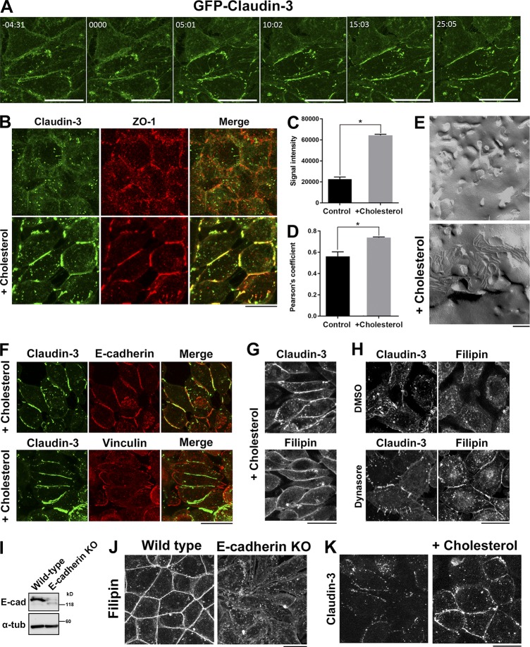
Addition of cholesterol to the PM induces TJ strand formation in α-catenin–KO cells. (A) Time-lapse imaging of α-catenin–KO EpH4 cells expressing GFP–claudin-3. At time 0, 75 mM cholesterol-saturated MβCD was added to the medium to restore the level of cholesterol in the PM. (B) α-Catenin–KO EpH4 cells were treated with PBS (control) or 75 mM cholesterol-saturated MβCD, fixed, and costained with an anti–claudin-3 pAb (green) and an anti–ZO-1 mAb (red). (C) Quantification of the signal intensity of claudin-3 at cell–cell contact areas in α-catenin–KO EpH4 cells before and after loading of cholesterol in the PM. (D) Quantification of the colocalization of claudin-3 and ZO-1 in α-catenin–KO EpH4 cells before and after loading of cholesterol in the PM. The degree of colocalization between claudin-3 and ZO-1 was calculated using ImageJ FIJI software. The value of Pearson’s coefficient of two signals were quantitated. Error bars show SD calculated based on four independent experiments (Student’s t test, *, P < 0.05). (E) Freeze-fracture EM images of TJ strands in α-catenin–KO EpH4 cells treated with PBS (control, top) or 75 mM cholesterol-saturated MβCD (bottom) for 30 min. (F) α-Catenin–KO EpH4 cells were treated 75 mM cholesterol-saturated MβCD, fixed, and stained with an anti–claudin-3 pAb (green) together with an anti–E-cadherin mAb (red, top) or an antivinculin mAb (red, bottom). (G) α-Catenin–KO EpH4 cells expressing GFP–claudin-3 were treated with 75 mM cholesterol-saturated MβCD, fixed with 4% paraformaldehyde, and stained with 50 µg/ml filipin prepared in PBS. (H) α-Catenin–KO EpH4 cells expressing GFP–claudin-3 were treated with DMSO (control, top) or 100 µM dynasore (bottom), fixed with 4% paraformaldehyde, and stained with 50 µg/ml filipin prepared in PBS. (I) Immunoblotting of whole-cell lysates of WT and E-cadherin–KO EpH4 cells with the indicated antibodies. (J) WT and E-cadherin–KO EpH4 cells were fixed with 4% paraformaldehyde and stained with 50 µg/ml filipin prepared in PBS. (K) E-cadherin–KO EpH4 cells were treated with PBS (control) or 75 mM cholesterol-saturated MβCD, fixed, and stained with an anti–claudin-3 pAb. Bars: (A, B, F–H, J, and K) 20 µm; (E) 200 nm.

