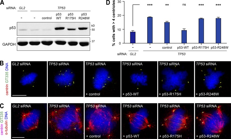
Figure 5.
The p53 hotspot mutations R175H and R248W deregulate centriole numbers in metaplasia. (A–D) Metaplasia cells depleted of endogenous p53 (TP53) were transfected with WT p53, p53-R175H, p53-R248W, or the empty plasmid (control). Metaplasia cells transfected with control siRNA (GL2) or siRNA against endogenous p53 (TP53) alone were also analyzed. (A) Protein levels were assessed by WB. GAPDH was used as a loading control. (B–D) Cells were stained for centrioles (centrin and GT335) and DNA (B) or centrioles, microtubules (α-tubulin), and DNA (C). (B and C) Representative images are shown. Bars, 10 µm. (D) Quantification of mitotic cells with centriole amplification. n ≥ 100/condition/experiment. Error bars show means ± SEM of two independent experiments. **, P < 0.01; ***, P < 0.001 (ANOVA).

