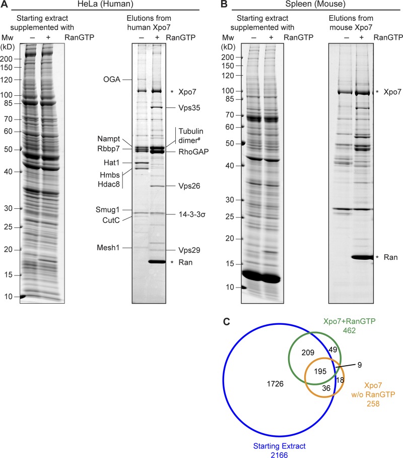
Figure 1.
Identification of novel Xpo7 binders. (A) Xpo7 tagged with the ED domains of protein A was immobilized on anti–protein A beads and incubated with 2 ml hypotonic HeLa extract (Abmayr et al., 2006) in the absence or presence of 5 µM RanGTP (Q69L ΔC terminus mutant). 1/2,000 of the starting extracts and 1/10 of the bound fractions were analyzed by SDS-PAGE followed by Coomassie staining. Indicated bands were identified by MS. Tubulin dimer# denotes tubulin α 4A and tubulin β 4B as the predominant forms. (B) Xpo7 affinity chromatography was performed as in A, but an extract from mouse spleen was used as starting material. Mw, molecular weight. (C) Starting extract (–Ran), Xpo7–RanGTP, and Xpo7 without Ran samples in B were analyzed by MS. The Venn diagram represents the number of identified unique proteins.

