Figure 1.
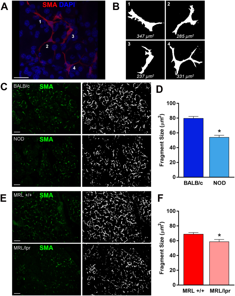
Determination of MEC size in healthy and diseased lacrimal glands. (A) Lacrimal glands from BALB/c mice were stained with an antibody against α-smooth muscle actin (SMA) and imaged by confocal microscopy to visualize whole MECs. (B) ImageJ was used to create masks of MECs in the normal lacrimal gland to generate the representative sizes. The same method was then used to compare MEC size, using SMA staining (green; C,E) in lacrimal glands of diseased and healthy mice. MEC staining was masked using ImageJ (white; C,E) and quantified to determine the average size of the stained fragments to compare NOD and BALB/c (C,D) or MRL/lpr and MRL +/+ (E,F). Star denotes statistically significant difference compared to control; n = 6 for MRL/lpr mice and n = 5 for all other groups; scale bar represents 25 µm (A) and 50 µm (C,E).

