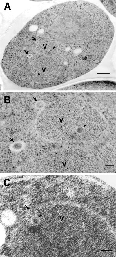
Figure 9.
Electron micrographs of sec12 under starvation conditions. sec12 mutant cells were cultured in SD(-N) containing PMSF at 37°C for 2.5 h and prepared for electron microscopy as described in MATERIALS AND METHODS. Higher magnification images are shown in B and C. Arrows, Cvt vesicle; arrowheads, Cvt body; V, vacuole. Bars, 500 nm (A); 100 nm (B and C).

