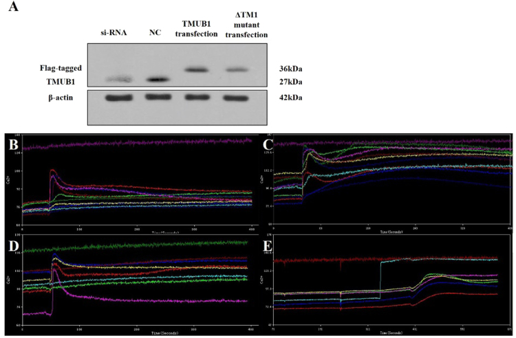
Figure 4.
The impact of TMUB1 on [Ca2+]i in BRL-3A hepatocytes. (A) The effects of transfection of full-length TMUB1, its mutants and siRNA were tested by Westernblotting. BRL-3A hepatocytes were prepped for experiments on day 2 when the cells reached 50–60% confluence; the cells were then transfected with pcDNA-TMUB1-Flag, pcDNA-ΔTM1-Flag, pcDNA-control and Tmub1-siRNA. (B–E) Measurement of [Ca2+]i levels. The cells were harvested and detached with 0.25% trypsin into a cell suspension 72 hours after transfection. Images were obtained from five randomly selected fields, and at least seven cells were observed per visual field (indicated by the different colored lines). The average peak values of [Ca2+]i in BRL-3A hepatocytes transfected with pcDNA-control (B), Tmub1-siRNA (C), pcDNA-ΔTM1-Flag (D) and pcDNA-TMUB1-Flag (E) were98.3 ± 1.5 nm, 128.1 ± 3.0 nm, 103.7 ± 3.5 nm, respectively, and nearly no peak wave was observed at baseline (the average baseline was 75.6 ± 3.5). TMUB1 knockout significantly increased [Ca2+]i and lasted for a long time, while overexpression of TMUB1 significantly inhibited [Ca2+]i. Furthermore, when the interaction of TMUB1 andCAML was disturbed by deletion of the binding site (ΔTM1), [Ca2+]i in the ΔTM1 group exhibited no significant changes compared with that in the control group (F = 268.3, P < 0.01, ANOVA between the four groups and P = 0.18, LSD between the ΔTM1 group and control group).

