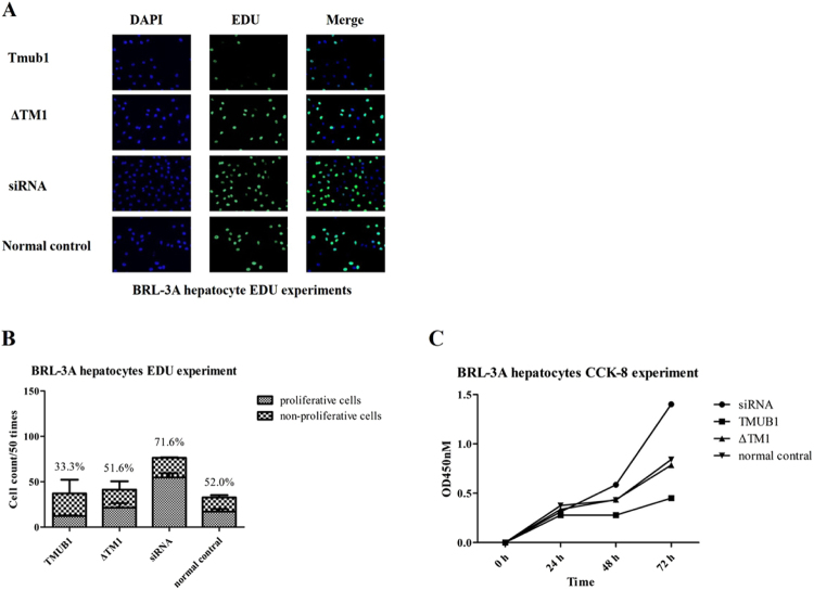
Figure 7.
TMUB1 inhibited BRL-3A hepatocyte proliferation through aninteraction with CAML (A,B). EDU experiments in BRL-3A hepatocytes. Hepatocytes transfected with TMUB1 had the fewest cells and lowest number of proliferative cells in every well. By contrast, TMUB1 knockout resulted in significantly more proliferative cells. When the interaction between TMUB1 and CAML was disturbed, the cell proliferation rates in the ΔTM1 group were nearly the same as those in the normal control group (X2 = 53.386, P < 0.01, chi-square test between four groups and X2 = 1.906, P = 0.107, between the ΔTM1 group and control group. The transfection test is shown in Fig. 6H). C. CCK-8 experiment in BRL-3A hepatocytes. Absorbance was not significantly different at 0 and 24 hours (F = 1.661, P = 0.252, ANOVA between four groups) but was significantly different at 48 (F = 9.690, P < 0.01, ANOVA between four groups) and 72 hours (F = 8.934, P < 0.01, ANOVA between four groups). When the interaction between TMUB1 and CAML was disturbed, the absorbance in the ΔTM1 group was almost the same as that in the normal control group at 24 hours (P = 0.438, LSD between the ΔTM1 group and control group), 48 hours (P = 0.941, LSD between the ΔTM1 group and control group) and 72 hours (P = 0.769, LSD between the ΔTM1 group and control group).

