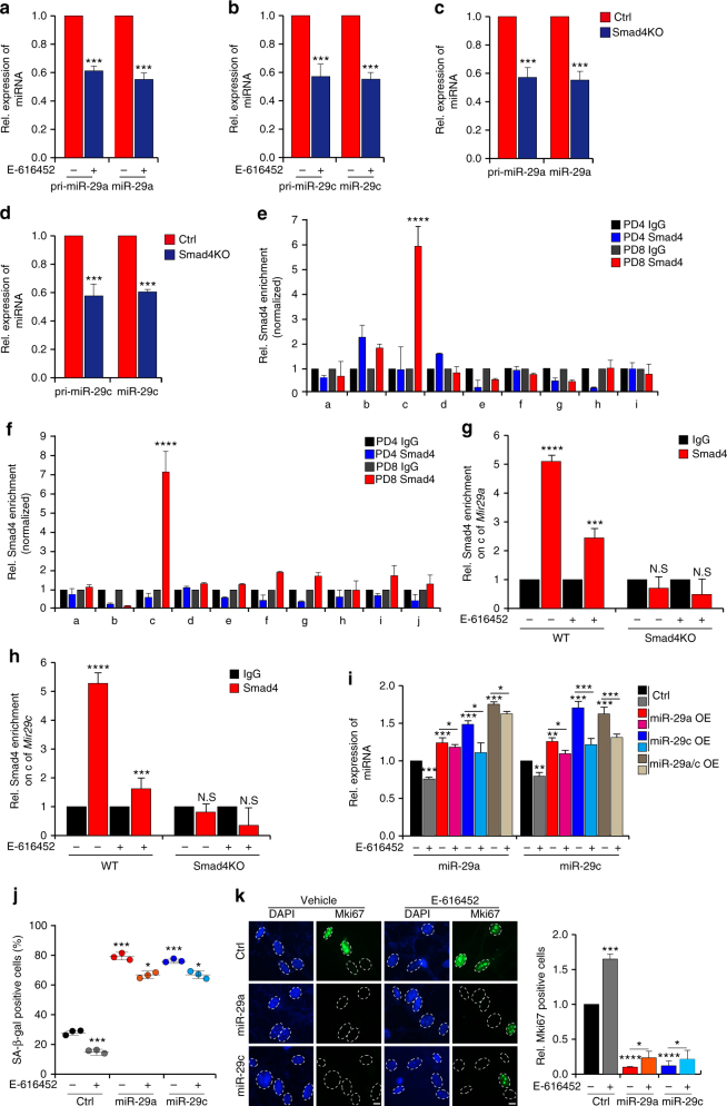
Fig. 3.
TGF-β signaling regulates miR-29 expression in a Smad-dependent manner during MEFs senescence. a–d Quantitative real-time PCR for pri-miR-29 and miR-29 in MEFs incubated with E-616452 (a, b) or Smad4 knockout (Smad4KO) MEFs (c, d). e, f Chromatin immunoprecipitation-qPCR (ChIP-qPCR) assays of Smad4 from PD4 and PD8 MEFs. The x-axis represents the primer positons of the predicted promoters of Mir29a (e) and Mir29c (f). g, h Smad4 enrichment on Mir29a (g) and Mir29c (h) was measured in inhibitor-treated MEFs and Smad4KO MEFs. i MEFs with lentivirus-mediated expression of miR-29 were incubated with E-616452. Real-time PCR was used to measure miR-29 expression. j Diagrams showing SA-β-gal staining of MEFs collected from i. k Immunofluorescent staining showing the Mki67 signals in MEFs with the same treatment as those shown in i. Scale bar, 5 μm. The error bars represent the s.d obtained from triplicate independent experiments. Two-tailed unpaired Student’s t-tests were performed, **p < 0.01, ***p < 0.001

