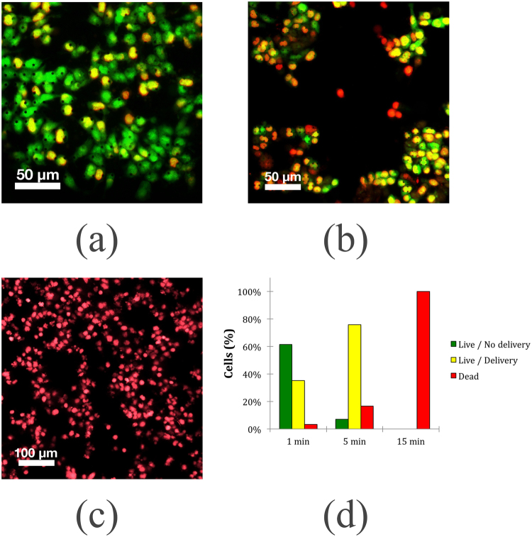
Figure 7.
Fluorescence imaging of calcein AM and EthD-1 stained HCT 116 cells after application of a magnetic field of 360 Oe and 425 kHz for three different times. (a) 1 minute. (b) 5 minutes. (c) 15 minutes. For each time point, three independent groups of cells were grown for 24 hours and then stained and imaged. A treatment time of 1 minute and 5 minutes showed delivery efficiencies of 35% and 75%, respectively. Cell death of the entire population was observed for the group of cells placed under a field for 15 minutes.

