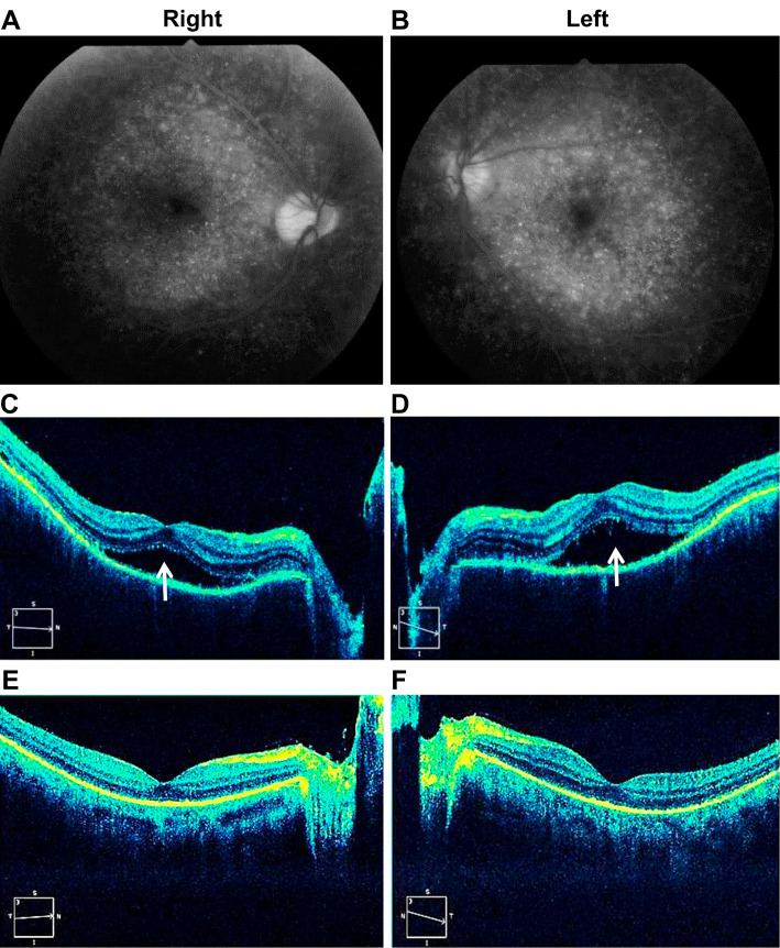
Figure 2.
Fluorescein fundus photography and optical coherence tomography. A, B: Fluorescein fundus photography shows diffuse multiple early hyperfluorescent points on hospital day 4. C, D: Optical coherence tomography shows serous retinal detachments (arrows) on hospital day 4. E, F: Optical coherence tomography shows that the bilateral serous retinal detachments diminished on hospital day 16.

