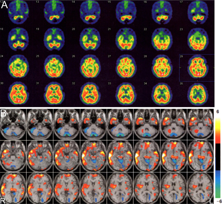
Figure 2.
99mTc-ECD SPECT of the brain (A) showed no apparent hypoperfusion in the cerebellum. Hyperperfusion was suspected in the bilateral lenticular nuclei and right temporal lobe. The two-tailed view display in eZIS (B) demonstrated regions showing a Z-score of ≤-2 in the bilateral cerebellar hemisphere and the upper portion of the cerebellar vermis. The hypoperfusion of the bilateral cerebellar hemisphere might have been associated with the arachnoid cyst in the posterior fossa. In contrast, there were regions showing a Z-score of ≥2 in the midbrain, right lenticular nucleus, bilateral thalamus, and bilateral frontotemporal lobes.

