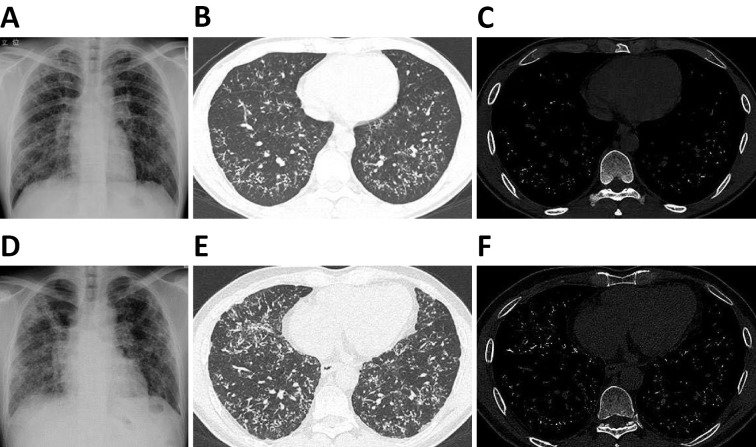
Figure 2.
Chest X-ray (A), CT image with lung window (B), and CT image with bone window (C) at 33 years of age. Chest X-ray (D), CT image with lung window (E), and CT image with bone window (F) at 46 years of age. Chest X-ray showed bilateral ground-glass opacity, and CT showed high-density nodules bilaterally in the lower lobes. Compared with the images at 33 years of age, the bilateral ground-glass opacity on chest X-ray had progressed, and the number of high-density nodules on CT had increased significantly at 46 years of age.

