Fig. 2.
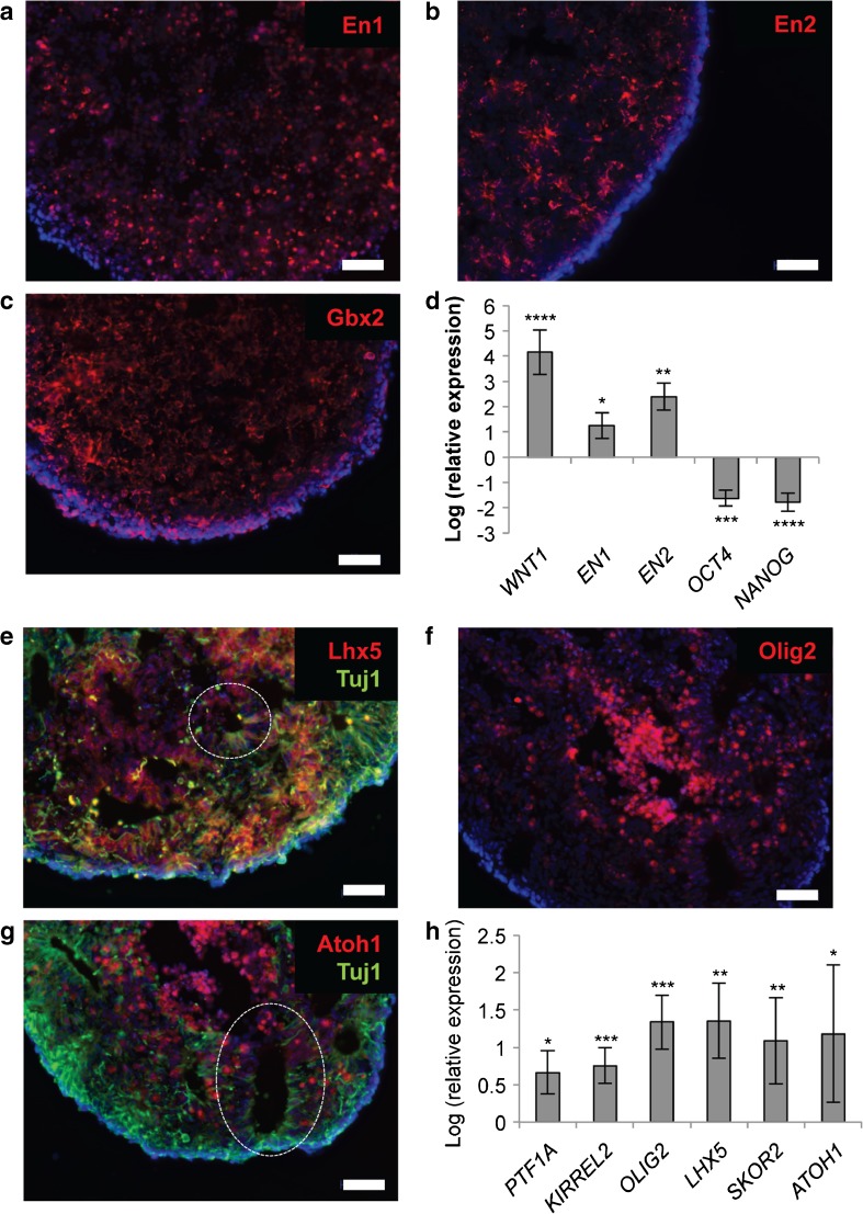
Generation of cerebellar progenitors from hiPSCs. After 21 days in suspension culture, hiPSC aggregates express hindbrain-specific transcription factors En1 (a), En2 (b) and Gbx2 (c), and the isthmic organiser factor WNT1, and show suppression of the pluripotency genes OCT4 and NANOG (d). By day 35, subpopulations of cells within hiPSC aggregates express the early neuronal marker Tuj1 (βIII-tubulin), as well as Purkinje cell precursor markers Lhx5 (e), Olig2 (f) and the granule cell marker Atoh1 (g). Expression of the ventricular zone (GABAergic) marker PTF1A, and the rhombic lip (glutamatergic) marker ATOH1, as well as additional Purkinje cell markers KIRREL2 and SKOR2, was confirmed by qPCR (h). Nuclei are stained with DAPI (blue). Examples of neural rosettes are indicated by a dashed line. Scale bar 50 μm. Gene expression is shown relative to hiPSCs (set to zero), normalised to β-actin. Results are representative of five biological replicates, except in the case of PTF1A, where n=2; error bars represent SD. *p < 0.05; **p < 0.01; ***p < 0.001; ****p < 0.0001

