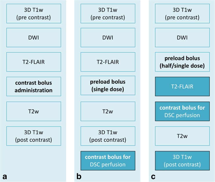
Fig. 3.
Three possible options for a glioma imaging protocol in clinical practice based on the EORTC-NBTS protocol (a), with the addition of DSC perfusion imaging (b, c). Option C has the advantage over option B that it has double the contrast dose for post-contrast T1w imaging. Option B may be preferred if non-contrast enhanced T2-FLAIR is desired. Please see Ellingson et al. [9] for further considerations and vendor-specific sequence details on structural and diffusion-weighted imaging. The moment of contrast administration is indicated in bold

