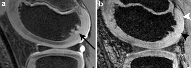
Fig. 6.
Ossification disturbance in the posterior part of the lateral femoral condyle. a IMw image with irregularities of the ossification centre (black arrow; see description in text). b SWI: the epiphyseal cartilage directly overlying the lesion shows a reduced density of vessels (black arrowhead, grade 0) while more proximally some vessels can be delineated faintly (white arrowhead). Also compare with the anterior part of the femoral condyle

