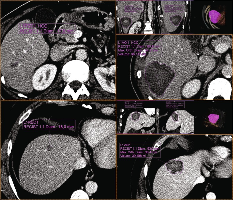
Fig. 2.
Left Preprocedural portal venous phase contrast enhanced CT images of HCC in segment 6 (top) and CRLM in segment 4 (bottom) in two patients who had not received systemic therapy or transarterial (chemo)embolization. Using MWA device B, 96 kJ (100 W for 8:00 min × 2) and 96 kJ (100 W for 6:00 min, 100 W for 10:00 min) were applied to the HCC and CRLM, respectively, with 16 mm and 14 mm between the two positions of the ablation center of the antenna, so overlap was approximately similar. Right Resulting ablation zones after segmentation on the 1-week follow-up portal venous phase contrast-enhanced CT images using the MM Oncology package (syngo.via; Siemens, Erlangen, Germany), with ablation zone volumes of 96 mL and 39 mL, resulting in energy deposition ratios of 1.00 mL/kJ and 0.41 mL/kJ for HCC and CRLM, respectively. After 6 months of follow-up, the HCC showed no sign of recurrence, whereas a PET scan of the CRLM showed activity at the dorsal side of the ablation zone, for which re-ablation was performed

