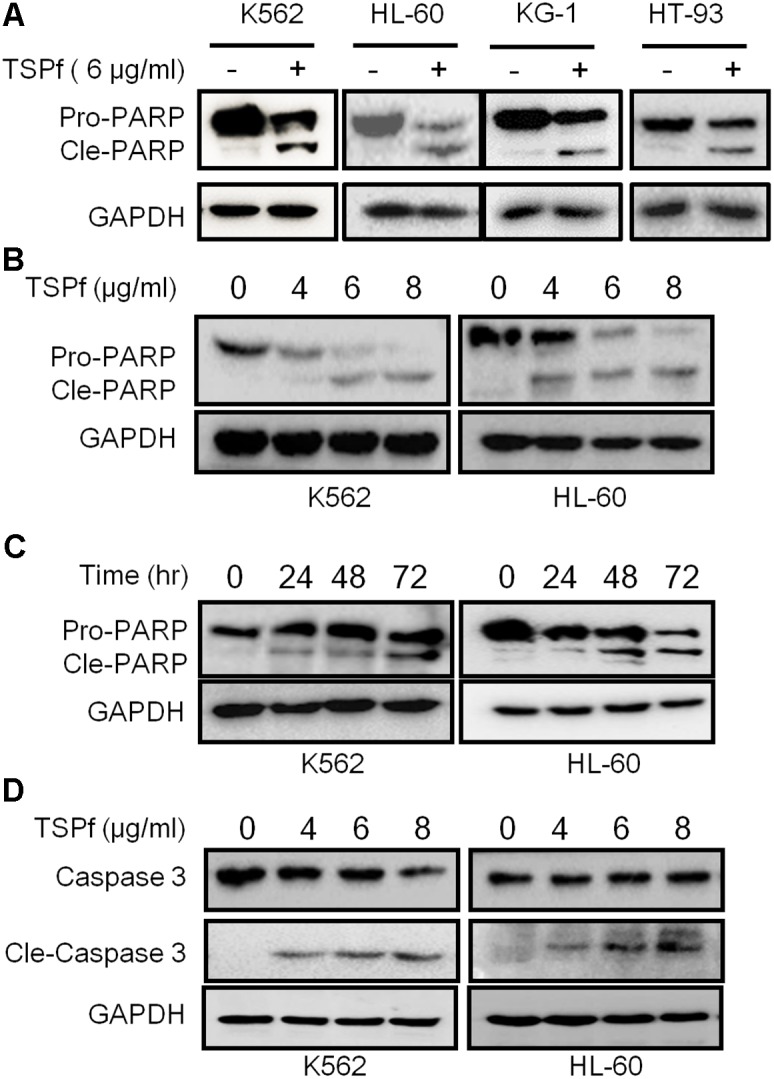
FIGURE 4.

TSPf induces leukemia cell apoptosis. (A) Leukemia cell lines were treated with 6 μg/ml TSPf or DMSO for 24 h followed by lysate preparation and immunoblotting analysis against PARP and GAPDH. (B,C) K562 and HL-60 cells were treated with TSPf at indicated concentrations (B) for 24 h or 4 μg/ml of TSPf for indicated periods (C) followed by analysis for PARP. GAPDH was used as a loading control. (D) K562 and HL-60 cells were treated with TSPf at indicated concentrations for 24 h followed by immunoblotting assays against caspase 3. GAPDH was used as an internal loading control.
