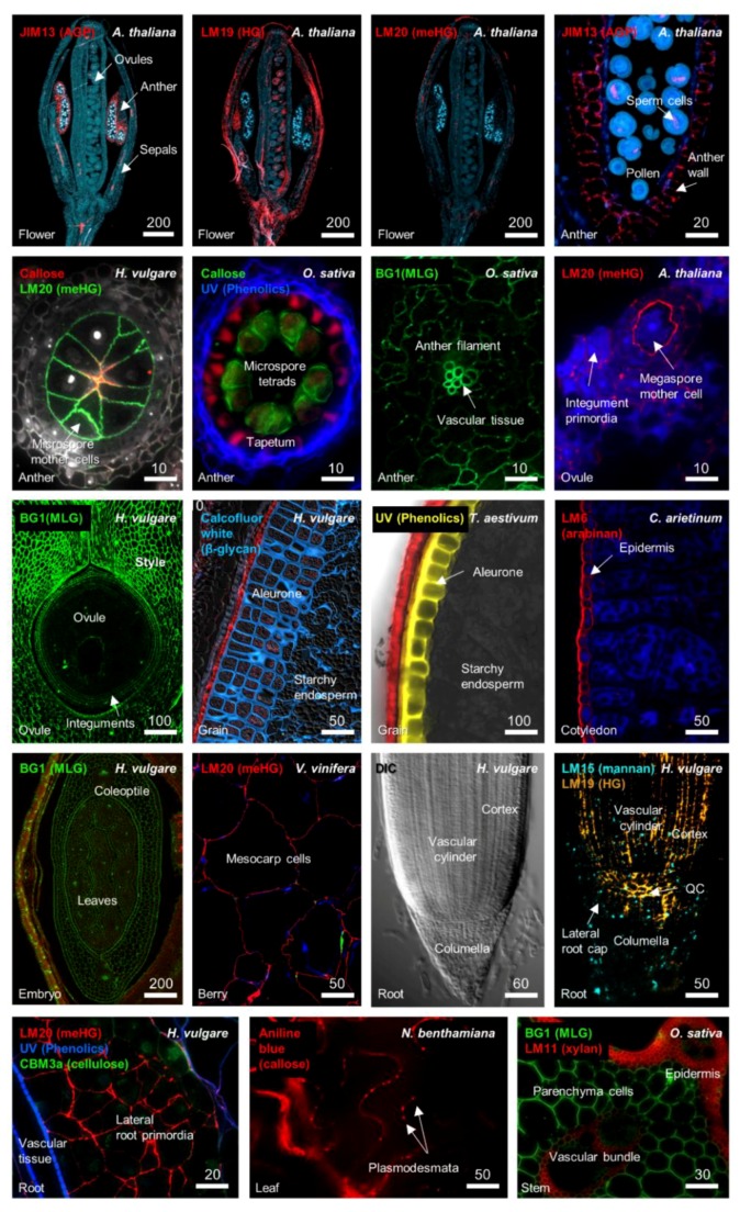
Figure 1.
Detection of different cell wall components in distinct tissues of Arabidopsis thaliana, Hordeum vulgare (barley), Oryza sativa (rice), Cicer arietinum (chickpea), Vitis vinifera (grape), Nicotiana benthamiana (tobacco), and Triticum aestivum (bread wheat). The tissue origin of each section is indicated at the bottom left of each panel. The antibody or stain is indicated at the top left of each panel. Labelling of polymers was achieved through the use of diverse antibodies including BG1 (1,3;1,4-β-glucan), JIM13 (arabinogalactan proteins, AGP), LM19 (homogalacturonan, HG), LM20 (methylesterified homogalacturonan, meHG), callose (1,3-β-glucan), LM15 (mannan), LM6 (arabinan), LM11 (arabinoxylan), and CBM3a (cellulose), or stains such as aniline blue (1,3-β-glucan) and Calcofluor White (β-glycan), or UV autofluorescence. Differential contrast (DIC) microscopy was used to image the barley root tip and is shown as a reference for the adjoining immunolabelled sample. Images were generated for this review, but further details can be found in previous studies [23,29,30,31,32]. Scale bar dimensions are shown in µm.

