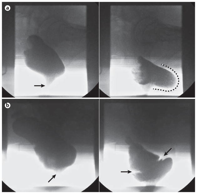
Figure 5.
Representative barium defecography images. a | A significant rectocele; the left panel shows a lateral view of the rectum at rest, opacified by barium neostool with the anal canal closed (arrow). The right panel clearly demonstrates a large retaining rectocele at end evacuation (extent of anterior bulging highlighted by dashed line) b) Obstructing full thickness intussusception; the left panel shows a lateral view of the rectum at rest, with the anal canal closed (arrow). The right panel shows an image at mid evacuation with clear invagination of the mid rectum (between arrows) secondary to a full thickness rectal intussusception; this is causing occlusion of the distal rectal lumen with retention of neostool proximal to this.

