Figure 4.
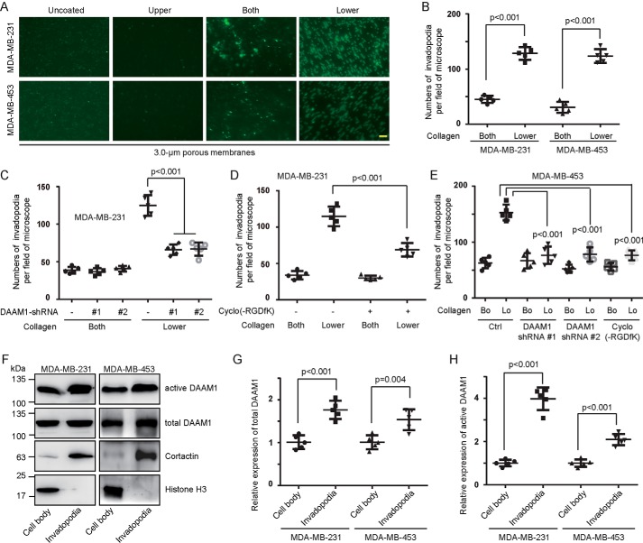
DAAM1 activation is essential for collagen-induced invadopodia extension. A and B, type IV collagen triggered invadopodia extension of MDA-MB-231 and MDA-MB-453 cells. MDA-MB-231 or MDA-MB-453 cells were examined for invadopodia extension for 6 h in 3.0-μm porous Boyden chamber membranes coated with vehicle (Uncoated) or type IV collagen on upper sides (Upper), both sides (Both), or lower sides (Lower). Invadopodia on the lower sides of the membrane were stained with cortactin antibodies and counted per field of the microscope. Bar, 10 μm. Objective lens, magnification, ×40; numerical aperture, 0.95. C, DAAM1 silence significantly inhibited collagen-induced invadopodia extension. Stable DAAM1 knockdown MDA-MB-231 cells (DAAM1 shRNA #1 and #2) or control cells were allowed to extend invadopodia for 6 h. The number of extended invadopodia was determined by 3.0-μm porous Boyden chamber assays and counted per field of the microscope. D, cyclo(-RGDfK) suppressed collagen-induced invadopodia extension. MDA-MB-231 cells were treated with 20 nmol/liter cyclo(-RGDfK) or vehicle and allowed to extend invadopodia for 6 h. The number of extended invadopodia was determined by 3.0-μm porous Boyden chamber assays and counted per field of the microscope. E, DAAM1 silence or cyclo(-RGDfK) significantly inhibited collagen-induced invadopodia extension. MDA-MB-453 cells transfected with DAAM1 shRNA or treated with cyclo(-RGDfK) were allowed to extend invadopodia for 6 h. The number of extended invadopodia was determined by 3.0-μm porous Boyden chamber assays and counted per field of the microscope. Bo, collagen coated on both sides. Lo, collagen coated on the lower sides. F–H, abundant active DAAM1 were located in invadopodia. MDA-MB-231 or MDA-MB-453 cells were seeded on 3.0-μm porous Boyden chamber membranes coated with collagen type IV on the lower sides. The invadopodia of MDA-MB-231 or MDA-MB-453 cells were allowed to extend toward collagen for 4 h. Cell bodies and invadopodia were separated as described under “Experimental procedures.” Cellular lysates were assayed for the active DAAM1 by a pulldown assay using a GST-RHOA as a bait. Error bars, S.D.

