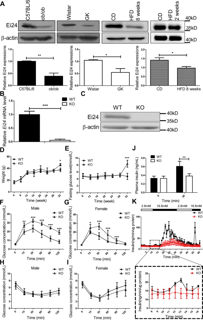
Figure 1.
Loss of Ei24 impairs glucose homeostasis. A, expression of Ei24 in islets from ob/ob mice, GK rats at 4 months of age, and C57BL/6 mice fed an HFD for 2 weeks and 8 weeks. β-Actin served as the loading control. CD, chow diet. B, total RNA was prepared from islets of WT and KO mice at 8 weeks of age. The transcription levels of Ei24 mRNA are normalized to β-actin mRNA. The results are representative of three individual experiments. C, Western blotting of Ei24 protein from isolated islets (300 islets/group) from WT and KO mice at 12 weeks of age. β-Actin served as the loading control. D, weight curves for WT and KO mice. The mean ± S.D. (error bars) of 15 mice is shown. E, concentration of the basic glucose curve for WT and KO mice. The mean ± S.D. of 10 mice is shown. F and G, glucose tolerance test (GTT) results of 10-week-old male (F) and female mice (G). Solid lines, WT mice (n = 8); dashed lines, KO mice (n = 8). H and I, ITT results of 10-week-old male (H) and female mice (I). Solid lines, WT mice (n = 8); dashed lines, KO mice (n = 8). J, in vivo GSIS detection in WT and KO mice. The results are representative of five replicates for each group. K, in vitro GSIS from isolated islets (70/group) of WT and KO mice by a fast digital perfusion system. The results are representative of three individual experiments. Data are expressed as the mean ± S.D. *, p < 0.05; **, p < 0.01; ***, p < 0.001.

