Figure 2.
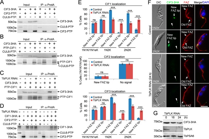
CIF3 interacts with CIF1 in a TbPLK-dependent manner. A, immunoprecipitation of CIF2–PTP was unable to pull down CIF3–3HA from trypanosome cell lysate. Cells expressing CIF2–PTP alone, CIF3–3HA alone, or both CUL6–PTP and CIF3–3HA served as the controls. B, immunoprecipitation of PTP–CIF1 was able to pull down CIF3–3HA from trypanosome cell lysate. Co-immunoprecipitation was carried out as above. CUL6–PTP was co-expressed with CIF3–3HA, and co-immunoprecipitation was carried out to rule out the possibility that the PTP tag can pull down CIF3–3HA. C, CIF3–CIF1 interaction depends on TbPLK. TbPLK RNAi was induced for 24 h. The asterisk indicates a degradation fragment of PTP–CIF1. D, CIF2–CIF1 interaction is independent of TbPLK. TbPLK RNAi was induced for 24 h. Cells co-expressing CUL6–PTP and CIF1–3HA were used as a control to show that the PTP tag was not able to pull down CIF1–3HA. The asterisk indicates a degradation fragment of CIF1–3HA. E, effect of TbPLK RNAi on the localization of CIF1, CIF2, and CIF3 during different cell cycle stages. Total numbers of cells counted are as follows. For CIF1 localization: 1N1K/1N1eK, 344 (control) and 334 (TbPLK RNAi); 1N2K, 167 (control) and 154 (TbPLK RNAi); 2N2K, 211 (control) and 256 (TbPLK RNAi). For CIF2 localization: 1N1K/1N1eK, 619 (control) and 310 (TbPLK RNAi). For CIF3 localization: 1N1K/1N1eK, 325 (control) and 308 (TbPLK RNAi); 1N2K, 154 (control) and 152 (TbPLK RNAi); 2N2K, 216 (control) and 220 (TbPLK RNAi). Error bars represented S.D. from three independent experiments. ***, p < 0.001; ns, no statistical significance. The label New FAZ tip+ indicates that CIF proteins are detected at the new FAZ tip, whereas the label New FAZ tip− indicates that CIF proteins are not detected at the new FAZ tip. F, localization of CIF3–3HA in noninduced control and TbPLK RNAi cells. Cells were co-immunostained with FITC-conjugated anti-HA mAb and anti-CC2D pAb to label CIF3–3HA and the FAZ, respectively. The white arrowheads indicate the weak CIF3 fluorescence signal at the old FAZ tip. Scale bar: 5 μm. G, CIF3 protein levels in control and TbPLK RNAi cells. TbPSA6 served as the loading control.

