Figure 4.
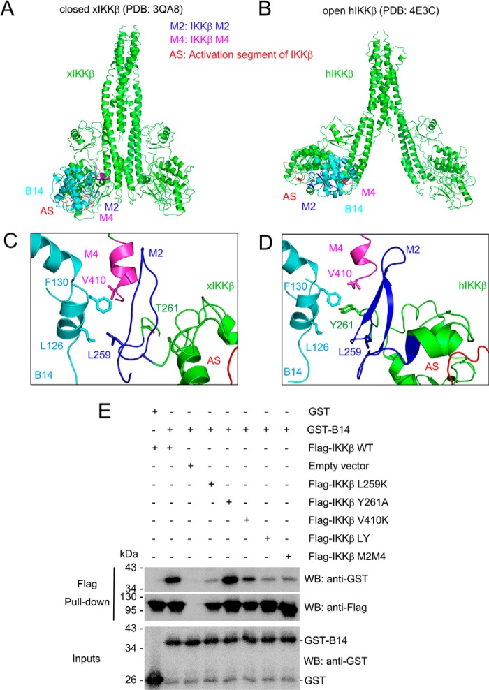
B14-IKKβ binding interface. A and B, models of B14/IKKβ complex structures. Closed Xenopus IKKβ (xIKKβ, PDB code 3QA8, A) and open human IKKβ (hIKKβ, PDB code 4E3C, B) structures are shown as ribbons and colored green. VACV B14 is shown as a ribbon and colored cyan. M2, M4, and the activation segment of IKKβ (AS) are colored blue, magenta, and red, respectively. C and D, B14-IKKβ binding interface for xIKKβ and hIKKβ, respectively. The key interacting residues are shown as sticks. E, GST-B14 was pulled down by human WT and mutant IKKβ. The empty vector was expressed as a control. LY refers to the L259K and Y261A IKKβ double mutant. The M2M4 IKKβ mutant contains both the M2 and M4 substitutions described in Fig. 3. WB, Western blot. The experiment was repeated three times with consistent results.

