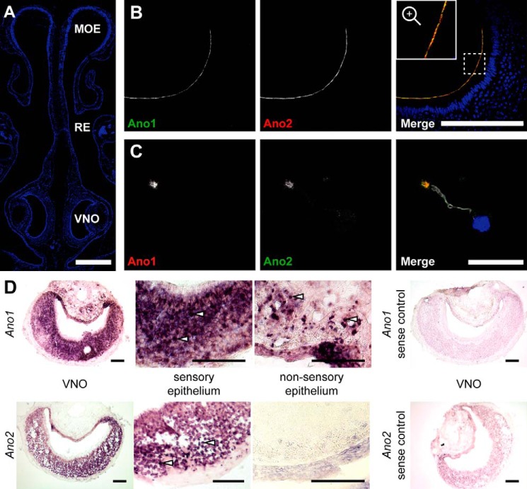
Figure 1.
Co-localization of Ano1 and Ano2 in vomeronasal sensory neurons. A, 4′,6-diamino-2-phenylindole-stained coronal section of the nose showing the morphology of the murine nasal cavity with MOE, respiratory epithelium (RE), and VNO. Bar, 500 μm. B, coronal sections of the VNO immunolabeled for Ano1 (green) and Ano2 (red, gpAno2_C1-3) at the apical border of the vomeronasal sensory epithelium. Bar, 200 μm. The region marked with a dashed square is magnified in the inset. C, immunostainings of isolated VSNs labeled for Ano1 (red), Ano2 (green, gpAno2_C1-3), and acetylated tubulin (white) at a single dendritic knob. Bar, 20 μm. The nuclei are colored blue in merged images. D, in situ hybridizations in coronal VNO slices using a probe against Ano1 or Ano2 with respective sense control hybridizations. Higher magnification images of the sensory and nonsensory epithelium are shown; see white arrows for examples of single cell bodies. All bars, 100 μm.

