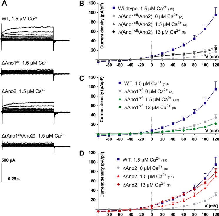
Figure 4.
Steady-state Ca2+-activated Cl− currents in vomeronasal sensory neurons. Steady-state Ca2+-activated Cl− currents of VSNs in acute slices from the VNO recorded in the whole-cell voltage clamp configuration are shown. Free intracellular Ca2+ concentrations in the patch pipette are indicated. A, example current traces at different voltage steps (−100 to 120 mV in steps of 20 mV); genotypes are indicated. B–D, current density–voltage plots of Ca2+-activated Cl− currents measured in different genotypes with either 0, 1.5, or 13 μm free Ca2+ in the patch pipette. The data from WT VSNs is plotted as reference. Numbers in parentheses indicate the number of measured cells. Mean current densities at respective voltage steps + S.E. (for clarity, shown only in positive direction).

