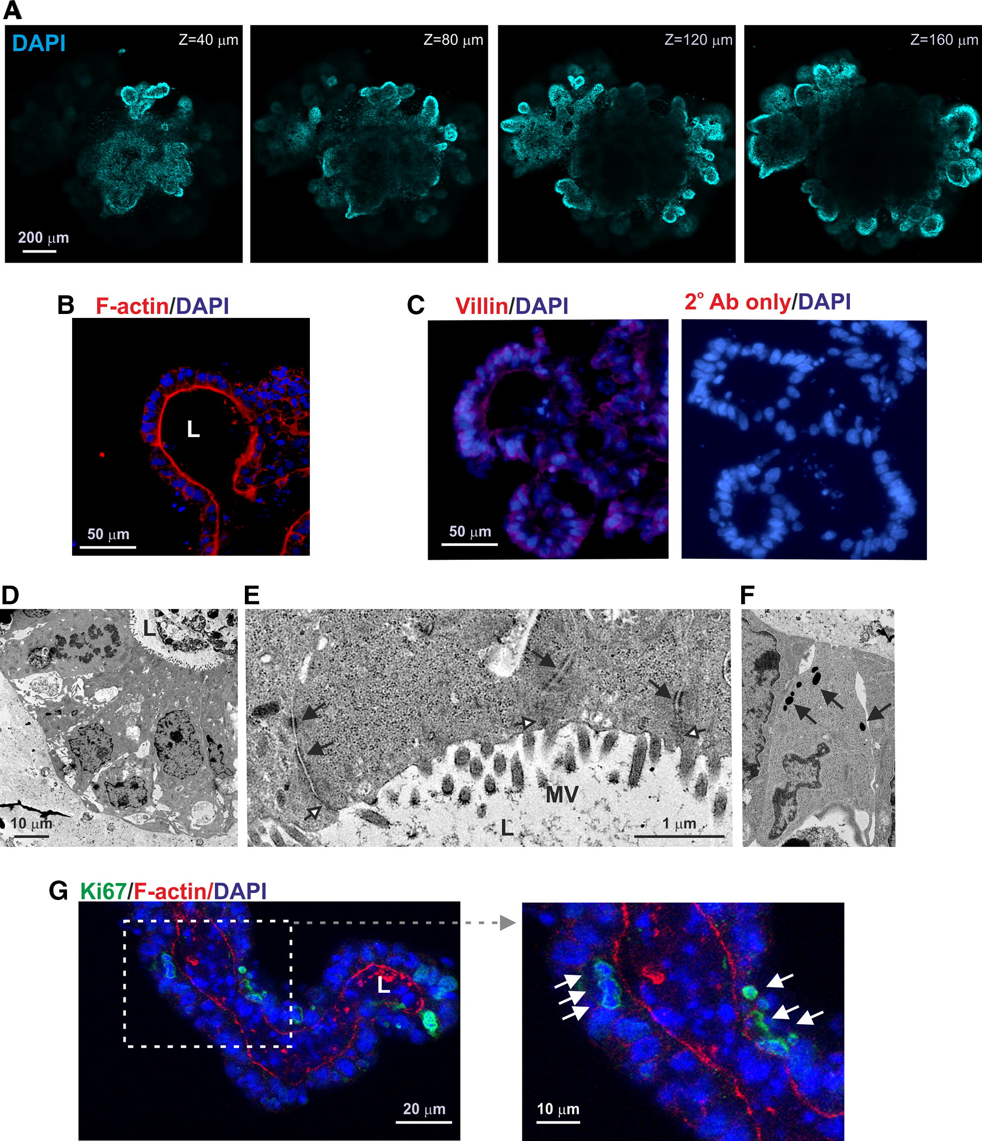
Figure 3.

Histological analysis of bovine enteroids. A Representative Z axis-projections of enteroids whole-mount stained to detect cell nuclei (DAPI, blue). B Detection of F-actin-expressing brush borders at the enteroid lumenal surface (L). C IHC analysis of villin expression in bovine enteroids. D–F, Ulstrastructural analyses of bovine enteroids. E Ultrastructural analyses shows the presence of microvilli (MV) on the apical surface of the enterocytes. The enterocytes are sealed by tight junctions (open arrows) and connected by desmosomes (closed arrows). F Detection of occasional cells containing dense cytoplasmic vesicles indicative of anti-microbial factor secreting Paneth cells. G IHC detect of abundant Ki67+ proliferating cells (green).
