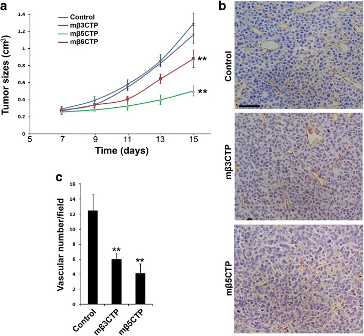
Fig. 3.
The antiangiogenic mβCTPs show antitumor activity. a 1.2 × 10⁶ of RM1 cancer cells were subcutaneously injected into BALB/c nude mice (n = 6). Starting on day 5, the formed tumor areas were subjected to treatment by local injection of 100 μl of the indicated mβCTP solution (50 μM at a final concentration) every other day. PBS alone was used as a control. Length (l) and width (w) of the formed tumors were measured, and tumor volumes (v) were calculated by using the following formula: v = 0.52(l × w2). b, c The tumor tissues were harvested on day 15 and subjected to IHC staining for CD31, a marker of vascular endothelial cells. Blood vessels in solid tumor tissues were quantified. Scale bar, 50 μm. The results were expressed as means ±SD; statistical significance was analyzed using Student’s t test (**, p < 0.01)

