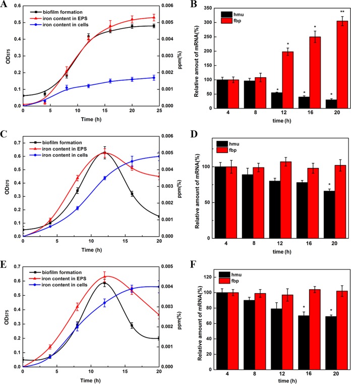
FIG 5.
Relationship between QS-dependent iron uptake systems and biofilm formation. (A) Development of wild-type biofilm formation and the iron content in the EPS and cells enveloped in the EPS. Biofilms were stained with crystal violet and quantified by their absorbance at 575 nm. The iron concentration was determined during biofilm development and is shown in parts per million. (B) RT-qPCR of hmu and fbp operons during biofilm formation of the wild-type strain. Data were analyzed using the 2−ΔΔCT method. (C) Development of ΔpdeI mutant biofilm formation and the iron content in the EPS and cells enveloped in the EPS. (D) RT-qPCR of hmu and fbp operons during biofilm formation of the ΔpdeI mutant. (E) Development of ΔpdeR mutant biofilm formation and the iron content in the EPS and cells enveloped in the EPS. (F) RT-qPCR of hmu and fbp operons during biofilm formation of the ΔpdeR mutant. Asterisks indicate a significant increase compared to the control during the same treatment period (*, P < 0.1; **, P < 0.05; ***, P < 0.01).

