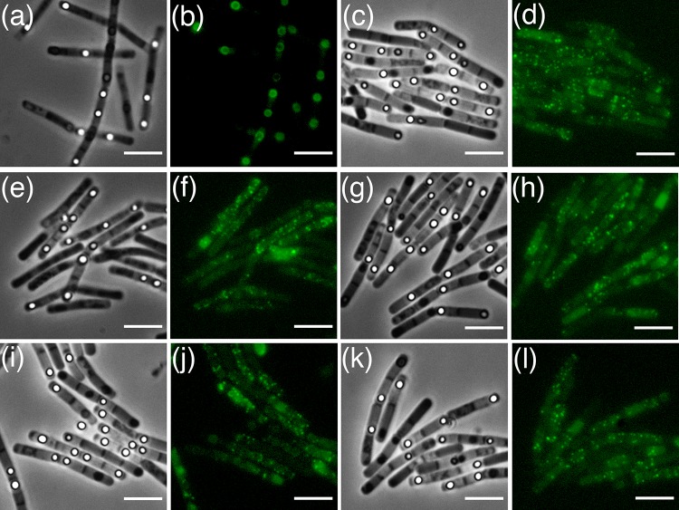
FIG 5.
Phase-contrast and fluorescence microscopy of sporulating B. cereus gerPA cells with plasmid-borne copies of gerPA-gfp (a and b), gerPB-gfp (c and d), gerPC-gfp (e and f), gerPD-gfp (g and h), gerPE-gfp (i and j), and gerPF-gfp (k and l). None of the remaining GerP proteins can localize around the developing forespore in the absence of GerPA. Scale bar = 5 μm.

